Rib Cage Muscles Diagram / Anatomytools Com Human Anatomy Human Anatomy For Artists Rib Cage Anatomy : Rib 2 is thinner and longer than rib 1 and has two articular facets on the head as normal.
Rib Cage Muscles Diagram / Anatomytools Com Human Anatomy Human Anatomy For Artists Rib Cage Anatomy : Rib 2 is thinner and longer than rib 1 and has two articular facets on the head as normal.. Start studying rib cage muscles. In humans, the rib cage, also known as the thoracic cage. Structure of a typical rib: So, let's learn the ribs so we can so what parts of the rib cage show up on the surface? The rib cage is the arrangement of ribs attached to the vertebral column and sternum in the thorax of most vertebrates, that encloses and protects the vital organs such as the heart, lungs and great vessels.
Further, there are two superior and two inferior processes meant for articulation with the neighbouring vertebra. The primary responsibilities of the ribcage involve protecting the thoracic visceral organs, enclosing the thoracic visceral organs, and is included in the general mechanics of the process of breathing. Target your rib muscles with specific exercises. When the upper arm is lifted away from the torso, the the thick outer edge is the anterior wall of the axillary (armpit) region. The rib cage has three important functions:
Back Pain And Slipped Rib from www.spineuniverse.com This post is about rib cage. Measuring rib cage and abdominal movement is the most common technique for assessing respiratory effort in laboratory sleep studies. Your ribs form a protective cage that encloses many of your delicate internal organs, such as your heart and lungs. Structure of a typical rib: In humans, the rib cage, also known as the thoracic cage. Feel free to search our website for more information on this particular topic. Anterior view of the lungs and ribcage in a transparent female torso stock illustration these pictures of this page are about:human anatomy rib cage muscles. Introduction to the structure of the ribcage and ribs:
Anterior view of the lungs and ribcage in a transparent female torso stock illustration these pictures of this page are about:human anatomy rib cage muscles.
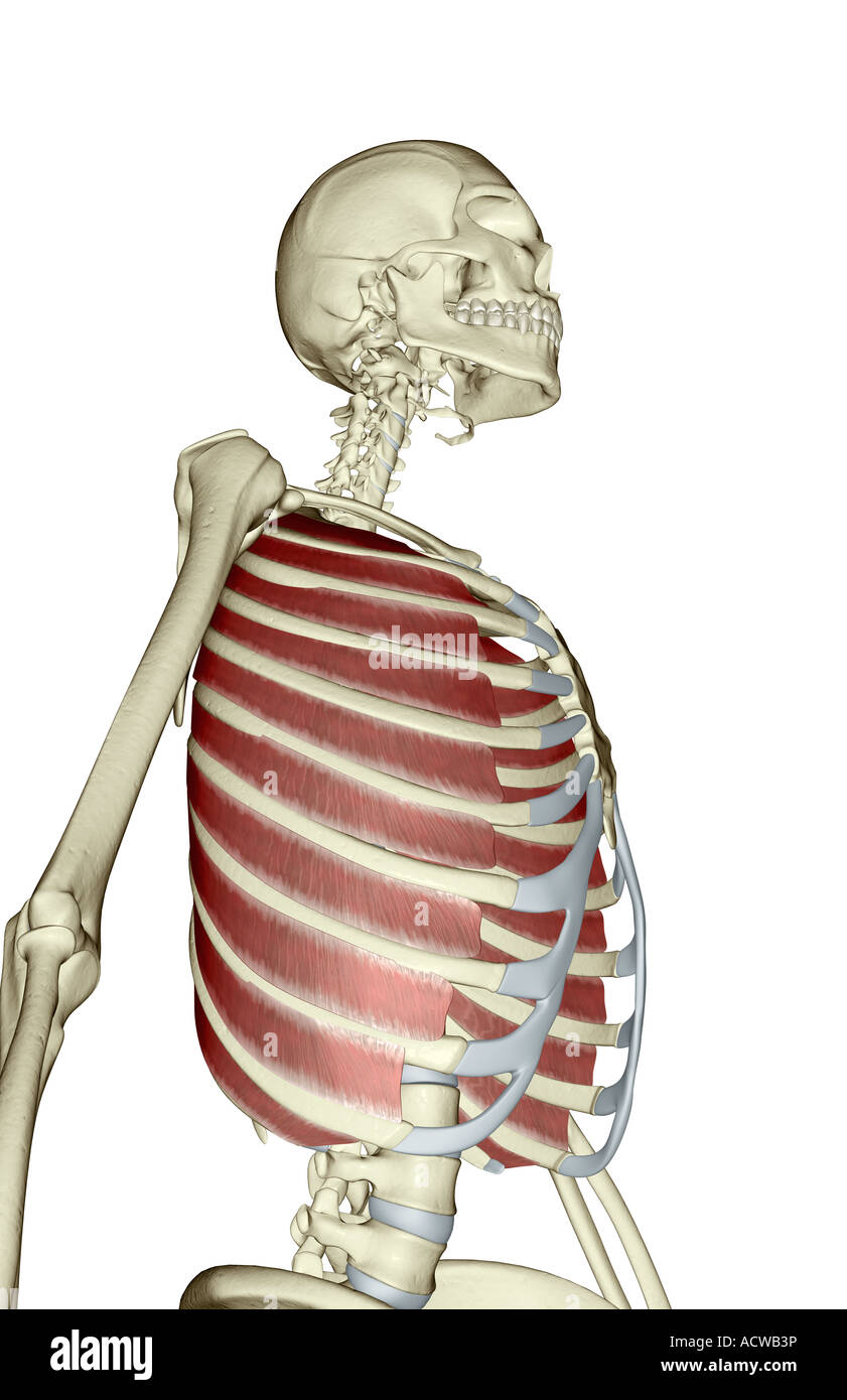
The other attachment of these muscles is usually considered to be either superior or inferior to the rib attachment. The other attachment of these muscles is usually considered to be either superior or inferior to the rib attachment. The following general rules regarding actions can be. Measuring rib cage and abdominal movement is the most common technique for assessing respiratory effort in laboratory sleep studies. Various skeletal muscles are attached to the rib cage. These rib muscles automatically get worked when you do bench presses, push ups and dips, but a few bonus exercises can help you really zero in for a more chiseled torso. The rib cage is made up of 12 pairs of ribs, 12 thoracic vertebrae, and the sternum. The accompanying diagram reveals the actions of the muscles in this pose. It provides a strong framework onto which the muscles of the shoulder girdle, chest, upper abdomen and back can attach. The rib cage is composed by sternum, costal cartilages, and ribs connected to the thoracic vertebrae. Each articulates with a thoracic vertebra. There are twelve (12) pairs of ribs and all articulate posteriorly with the thoracic vertebrae. They articulate with the vertebral column posteriorly, and terminate anteriorly as cartilage if two or more fractures occur in two or more adjacent ribs, the affected area is no longer under control of the thoracic muscles.
Your ribs form a protective cage that encloses many of your delicate internal organs, such as your heart and lungs. Skeletal muscles attached to the rib cage: The other attachment of these muscles is usually considered to be either superior or inferior to the rib attachment. It encloses and protects the heart and lungs. Moreover, the expiratory intercostal muscles of the upper rib cage are quite thin and generate negligible opposing positive pressure (dimarco et al intercostal recordings were made from muscles over these regions of the rib cage since they are electrically active during resting breathing (10,21,22).
Skeletal System Diagrams Human Body Anatomy Body Anatomy Human Ribs from i.pinimg.com Review the anatomical characteristics of the rib and ribcage in this interactive tutorial and test your knowledge in the quiz. On a muscular person when the muscles stretch, we see some of the lower. So what parts of the rib cage show up on the surface? During normal breathing, the major inspiratory muscles produce rib cage expansion and a downward movement of the diaphragm. These muscles may be located anteriorly, posteriorly, and/or laterally. The primary responsibilities of the ribcage involve protecting the thoracic visceral organs, enclosing the thoracic visceral organs, and is included in the general mechanics of the process of breathing. Some extend from above and draw the. Rib cage diagram with organs.
Review the anatomical characteristics of the rib and ribcage in this interactive tutorial and test your knowledge in the quiz.
It provides a strong framework onto which the muscles of the shoulder girdle, chest, upper abdomen and back can attach. The other attachment of these muscles is usually considered to be either superior or inferior to the rib attachment. The two muscles which comprise the intermediate muscle group are the serratus posterior inferior, and the serratus posterior superior. The ribs joint as follows: Review the anatomical characteristics of the rib and ribcage in this interactive tutorial and test your knowledge in the quiz. The muscle fibers pull across the rib cage and converge to attach on the humerus (upper arm bone). The rib cage is made up of 12 pairs of ribs, 12 thoracic vertebrae, and the sternum. The function of the rib cage is to filter the blood it receives, processing the blood. Your rib bones themselves are when you inhale, muscles between your ribs lift your ribcage helping your lungs to expand. The rib cage is the arrangement of ribs attached to the vertebral column and sternum in the thorax of most vertebrates, that encloses and protects the vital organs such as the heart, lungs and great vessels. These muscles may be located anteriorly, posteriorly, and/or laterally. These bony projections are used for attachment of muscles. The other attachment of these muscles is usually considered to be either superior or inferior to the rib attachment.
The ribs joint as follows: Posted on december 22, 2018december 22, 2018. When you exhale, the rib cage moves down again, squeezing the air. The last diagram shows how the ribs are connected to the vertebral column or spine. During normal breathing, the major inspiratory muscles produce rib cage expansion and a downward movement of the diaphragm.
External Intercostal Muscles Stock Photo Alamy from c8.alamy.com Posted on december 22, 2018december 22, 2018. This post is about rib cage. The rib cage labeled diagram. The ribs joint as follows: Rib cage diagram this summary post is displaying rib cage diagram. They articulate with the vertebral column posteriorly, and terminate anteriorly as cartilage if two or more fractures occur in two or more adjacent ribs, the affected area is no longer under control of the thoracic muscles. Start studying rib cage muscles. Muscles of thorax, upper extremities, back and diaphragm are given connection by this cage.
The other attachment of these muscles is usually considered to be either superior or inferior to the rib attachment.
There is a printable worksheet available for download here so you can take the quiz with pen and paper. Muscles that move the rib cage attach to the rib cage. It is formed by the vertebral column, ribs, and sternum and encloses the heart and lungs. Some extend from above and draw the. Target your rib muscles with specific exercises. Anterior view of the lungs and ribcage in a transparent female torso stock illustration these pictures of this page are about:human anatomy rib cage muscles. It encloses and protects the heart and lungs. Rib cage diagram with organs. The rib cage has three important functions: Muscles of thorax, upper extremities, back and diaphragm are given connection by this cage. • raise rib cage for inhaling & depresses rib cage for exhaling. So, let's learn the ribs so we can so what parts of the rib cage show up on the surface? In humans, the rib cage, also known as the thoracic cage.
Diversitech condensate pump wiring diagram rib cage muscles. Muscles of thorax, upper extremities, back and diaphragm are given connection by this cage.


0 Komentar